UC Davis vets use near-infrared imaging to treat chylothorax
A technique pioneered at the UC Davis Veterinary Medical Teaching Hospital is helping advance surgery for a potentially deadly condition.
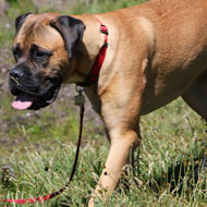
Near-infrared imaging has been used in the case of Atlas, a three-year-old male mastiff who initially presented with decreased energy levels and a respiratory rate that was increasing more than normal, even on the easiest of walks. His concerned owners took him to his vet who found he had a build up of a milky white fluid called chyle in his chest - a condition known as chylothorax, which can be deadly if not treated properly.
Atlas was referred to the specialists at the University of California Davis Veterinary Medical Teaching Hospital (UC Davis) and had almost 2,000 millilitres of the fluid, which leaks from the thoracic duct, removed from his chest. The removal of the fluid helped Atlas temporarily feel better but the cause of the chylothorax needed to be determined.
A number of things can cause the condition, including tumours in the chest cavity, heart disease, trauma to the chest causing damage to the thoracic ducts, heart worm disease, and idiopathic chylothorax in which no known underlying cause is identified. With Atlas, chest radiographs, an echocardiogram and a thoracic ultrasound ruled out any structural or functional abnormalities, and it was determined that he had idiopathic chylothorax.
Veterinarians discussed several options with Atlas' owners and they chose for him to have thoracic duct ligation. The procedure is often done via an open surgical approach but UC Davis offers thoracoscopic duct ligation - a minimally invasive surgery to correct the problem that offers the patient a quicker recovery and less discomfort and has also shown a similar success rate (80-85 per cent) compared to the typical more invasive thoracotomy.
This minimally invasive approach to chylothorax is used in human medicine and at a few veterinary schools but what sets UC Davis' approach apart is the use of intraoperative near-infrared imaging to make the lymphatic vessels more visible to the human eye during surgery.
Atlas underwent general anaesthesia for a CT lymphangiogram to outline the number and position of the thoracic ducts before Dr Phillipp Mayhew and Dr Michele Steffey proceeded with the ligation. The ducts that carry the chyle are very small and difficult to see under normal surgical illumination as they are buried in fat and other tissue.
The near infra-red approach, pioneered at UC Davis by Dr Steffey for this disease, uses different wavelengths of light, not visible to the human eye without special imaging systems. A special medical dye, that doesn't distort the appearance of tissues, is injected into a lymph node, carried to the ducts and is then visible when surgeons switch from a normal view to the near-infrared view through the scope, allowing them to see the position of the thoracic ducts clearly and enabling the dissection.
The near-infrared wavelengths travel through tissue better so surgeons can see the dye through several millimetres of tissue/ fat - where the naked eye can only see the surface of the tissue.
It's thought this technique may soon become the optimal protocol for these types of surgeries and in Atlas' case it enabled surgeons to easily locate his thoracic duct and stop the leakage. After two days of hospitalisation and oral pain medication he was back to his old self within a week, was given a clean bill of health at his one-month recheck and continues to do well nearly a year after the surgery.
Image courtesy of UC Davis


 The Greyhound Board of Great Britain has published new vaccination guidance, with all greyhounds registered from 1 January, 2027 required to have the L4 leptospirosis vaccination, rather than L2.